Top: The ultra high-field 7 Tesla MRI scanner at the LUMC is used in the study — Credits C.J. Gorter MRI Center
A new study on brain imaging by LEGend researchers, the WHIMAS study, has recently started data collection from memory clinic patients at the LUMC. The aim is to study novel cerebrovascular correlates of cognitive impairment and dementia on brain scans.
Dementia is a severe multifactorial, age-related disease resulting in memory loss and cognitive decline. It is often associated with reduced cerebrovascular function and cerebral white matter hyperintensities that can be detected with magnetic resonance imaging (MRI). A more in-depth analysis of these imaging markers is needed to further develop screening methods and future treatment strategies.

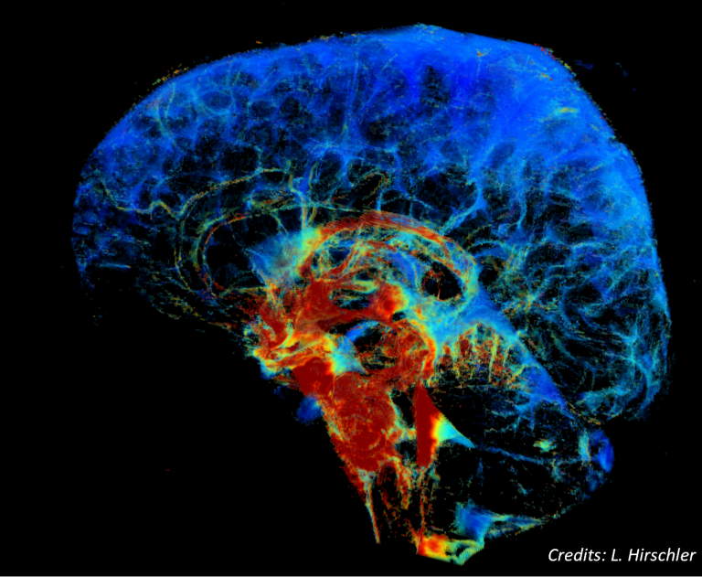
WHIMAS, or White Matter Hyperintensity Shape, is a study assessing structural cerebrovascular changes, but also the flow of cerebrospinal fluid that “cleans” the brain of waste and is thought to malfunction in several forms of dementia. Observations in these MRI scans can help to understand the complex brain changes in dementia.
The WHIMAS study is conducted by PhD candidates Jasmin Keller and Ingmar Eiling (PI: dr. Jeroen de Bresser). The study is a novel, close collaboration between the LUMC department of Radiology (Neuroradiology and the C.J. Gorter MRI Center) and the department of Internal Medicine. It involves multiple researchers of both departments. This research is supported by an LUMC theme grant and an Alzheimer Nederland grant. The study is expected to include 50 patients and will run throughout 2023.